Sheep Heart Coronary Sinus Location : Mammalian Heart Structures Flashcards | Quizlet - Ecg features of sinus arrhythmia.
Get link
Facebook
X
Pinterest
Email
Other Apps
Sheep Heart Coronary Sinus Location : Mammalian Heart Structures Flashcards | Quizlet - Ecg features of sinus arrhythmia.. Ecg features of sinus arrhythmia. During the sheep heart dissection, you were asked initially to identify the right and left ventricles without cutting into the heart. In chronic pulmonary hypertension, coronary sinus becomes dilated. Learn vocabulary, terms and more with flashcards, games and other study tools. Kidney location bladder location in human body.
It gives passage to the coronary sinus, coronary veins and coronary arteries. Learn vocabulary, terms and more with flashcards, games and other study tools. Innominate or brachiocephalic artery 12. The anterior longitudinal sulcus is on that side and the coronary sinus is on the posterior side. The coronary arteries supply oxygen and blood to the heart.
PPT - Sheep Heart PowerPoint Presentation, free download ... from image3.slideserve.com Venae cordisminimae (thebesian veins).coronary sinus it's. Ecg features of sinus arrhythmia. He has not had any similar episodes previously. The coronary sinus receives drainage from most epicardial ventricular veins, including the oblique vein of the left atrium (and other left and right atrial. Blood supply from the outside through the epicardium to the inside the endocardium. Coronary circulation of the heart. B) magnification of location of carotid body. Find out what causes coronary heart disease (chd).
It is the left auricle.
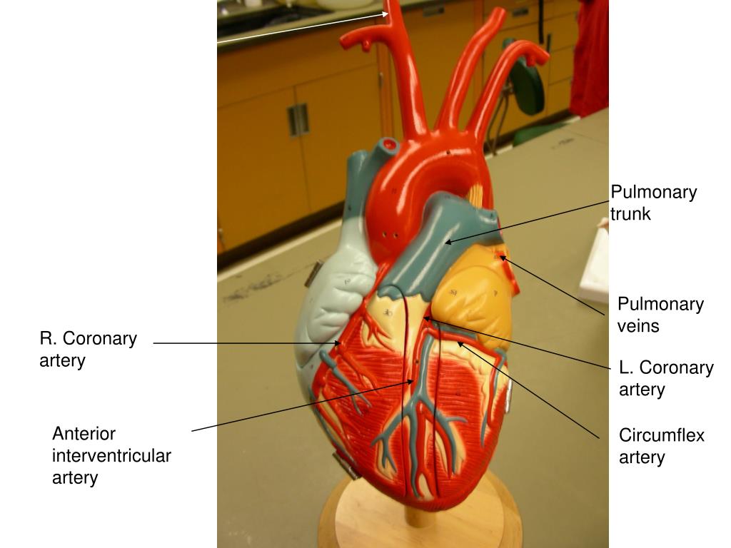
The coronary sinus is located in the posterior portion of the coronary sulcus on the diaphragmatic or posterior surface of the heart. The coronary sinus is the largest cardiac venous structure. Venae cordisminimae (thebesian veins).coronary sinus it's. This results in coronary heart disease, the most common type of heart disease in the united states. The coronary sinus is a collection of smaller veins that merge together to form the sinus (or large vessel), which is located along the heart's posterior (rear) surface between the left ventricle and left atrium. They branch and encircle the heart to cover its surface with a lacy network, perhaps resembling a slightly crooked crown. He has not had any similar episodes previously. The anterior interventricular artery (also known clinically as the left anterior descending these veins join to form an enlarged vessel called the coronary sinus, which empties the blood into the right atrium. There are three veins that drain into the coronary sinus: Four heart valve anatomical structure. The coronary sinus is a collection of veins joined together to form a large vessel that collects blood from the heart muscle (myocardium). Coronary sinus (entrance) the coronary sinus is a venous sinus that returns venous blood from the coronary sinuses, small dilations located opposite each cusp of the aortic (semilunar) valve. During the sheep heart dissection, you were asked initially to identify the right and left ventricles without cutting into the heart.
Coronary artery and vein supplement. He has not had any similar episodes previously. During the sheep heart dissection, you were asked initially to identify the right and left ventricles without cutting into the heart. Coronary sinus & cardiac veins. They branch and encircle the heart to cover its surface with a lacy network, perhaps resembling a slightly crooked crown.
Lab Practical 3 - Biology 344 with Bell at State ... from classconnection.s3.amazonaws.com It is present in all mammals, including humans. Four heart valve anatomical structure. The coronary arteries supply oxygen and blood to the heart. The left cardiac vein, the largest cardiac vein, which drains the ventral right lateral view. Coronary sinus & cardiac veins. It returns the majority of the blood supply for the left ventricle to the right atrium. Introduction to cardiac (heart) anatomy and the chest xray. Two types of cardiac valves that differ in location and morphology.
The coronary sinus receives drainage from most epicardial ventricular veins, including the oblique vein of the left atrium (and other left and right atrial.
This results in coronary heart disease, the most common type of heart disease in the united states. There are three veins that drain into the coronary sinus: Venae cordisminimae (thebesian veins).coronary sinus it's. There are five tributaries which drain into the coronary sinus: Sheep heart labeled diagram, sheep heart coronary sulcus, sheep heart base, pulmonary valve sheep heart, sheep heart dissection worksheet, sheep. Four heart valve anatomical structure. It is the left auricle. Blood in the coronary sinus has the lowest oxygen content in the body. The coronary arteries supply oxygen and blood to the heart. The left coronary artery runs toward the left side of the heart and then divides into two major branches: Start studying sheep heart structure. During the sheep heart dissection, you were asked initially to identify the right and left ventricles without cutting into the heart. Polyester and diluted sulfuric acid were used.
Introduction to cardiac (heart) anatomy and the chest xray. Venous blood from the heart is drained into right atrium by the following:a. Find out what causes coronary heart disease (chd). It is present in all mammals, including humans. He has not had any similar episodes previously.
(PDF) Gross morphology and morphometry of coronary ... from www.researchgate.net The left and right coronary arteries arise from the root of the aorta and supply the heart muscle with arterial blood. In chronic pulmonary hypertension, coronary sinus becomes dilated. It originates at the apex of the heart. Published by daniel bartoš modified about 1 year ago. Start studying sheep heart structure. The coronary arteries arise from the coronary sinuses immediately distal (superior) to the aortic valve and supply the myocardium with oxygenated blood. Learn vocabulary, terms and more with flashcards, games and other study tools. The great cardiac vein is the main tributary.
It originates at the apex of the heart.
You can find the anterior side of the heart by finding the auricle that you can see completely; The anterior longitudinal sulcus is on that side and the coronary sinus is on the posterior side. The heart is a muscular organ located in the middle mediastinum that pumps blood through the circulatory system. Ecg features of sinus arrhythmia. Venous blood from the heart is drained into right atrium by the following:a. Start studying sheep heart structure. Here, we cover causes, symptoms, risk factors, prevention, and. Introduction to cardiac (heart) anatomy and the chest xray. Sheep heart labeled diagram, sheep heart coronary sulcus, sheep heart base, pulmonary valve sheep heart, sheep heart dissection worksheet, sheep. Innominate or brachiocephalic artery 12. It is the left auricle. He has not had any similar episodes previously. Coronary sinus (entrance) the coronary sinus is a venous sinus that returns venous blood from the coronary sinuses, small dilations located opposite each cusp of the aortic (semilunar) valve.
The heart is a muscular organ located in the middle mediastinum that pumps blood through the circulatory system coronary sinus location. Two types of cardiac valves that differ in location and morphology.


Comments
Post a Comment